Heart Disease
The heart is a muscle pump located in the chest, which pumps blood around the body. It keeps pumping from a few weeks after conception until we die. There are a number of different conditions that can affect how the heart works.
What is the heart?
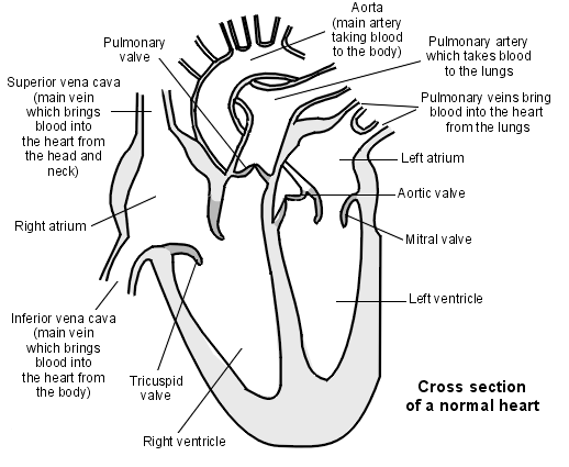
Normal heart detail

How does blood travel through the heart?
Blood that has been pumped around the body returns to the heart in the superior and inferior vena cava. It enters the first of the heart's four chambers, the right atrium. From there it is squeezed into the right ventricle which pumps the blood into the lung (pulmonary) circulation. In the lungs, blood picks up oxygen and returns to the left atrium of the heart. It passes through into the left ventricle, the most powerful part of the heart pump, which pumps the blood into the aorta (the main artery) and from there into the rest of the body.
Why the heart disease gender gap is costing women their lives
Coronary heart disease is a major cause of death among women worldwide, but it is often seen as ...
What can go wrong with the heart?
Any part of the heart can have a problem:
- The muscle wall of the heart (myocardium).
- The blood vessels supplying oxygen to the heart muscle (coronary arteries).
- The valves between the heart's four chambers.
- The conducting system that helps the heart to beat regularly and at the right speed.
- The covering of the heart (pericardium).
Heart disease symptoms
Chest pain is the most well-known symptom of heart disease. The typical pain which is caused by angina or a heart attack is described as a central crushing chest pain. It can sometimes cause a heavy aching in the left arm or in the neck or jaw. A heart attack also generally makes people feel sweaty, breathless and extremely unwell.
Other forms of heart disease often cause breathlessness and tiredness as not enough blood is getting pumped around the body to meet all its needs.
If there is a heart rhythm problem or the heart is beating fast then it may cause dizziness or a feeling of palpitations.
How are heart problems investigated?
If your doctor suspects you have a problem with your heart they will arrange some investigations.
Physical examination
The doctor will check your blood pressure and listen to your heart. Looking at other areas of the body as well may give them other clues about what is going on.
Blood tests
Your doctor may arrange for you to have some blood tests. These may:
- Check for anaemia which can make you feel breathless (full blood count).
- Check your cholesterol level (lipids).
- Check for diabetes (blood glucose or HbA1c).
- Check for heart failure (B natriuretic peptide, or BNP).
Electrocardiogram
Your doctor may arrange for you to have a heart tracing (electrocardiogram, or ECG). This will look at the electrical signals keeping your heart beating. This may be done by the practice nurse at your surgery or you may need to go to the hospital to have it done.
If you are experiencing the sensation of having a 'thumping' heart (palpitations) and your doctor suspects that your heart rhythm is sometimes irregular, they may want to arrange a 24-hour ECG.
Echocardiogram
If you have a heart murmur or if your doctor suspects heart failure, they may arrange for you to have an ultrasound scan of your heart (echocardiogram, or 'echo'). This looks at how the blood is flowing through the valves and the different chambers of the heart.
Coronary heart disease
This is what most people mean when they talk about heart disease. Coronary heart disease affects the coronary arteries, which are the blood vessels which supply the heart muscle. Sometimes it is called ischaemic heart disease. Ischaemic means blocked blood supply.
The arteries can become furred up with atheroma. Atheroma is made up of fatty deposits that form on the inside of blood vessels, which mean that blood does not flow normally through them.
Coronary heart disease includes:
- Angina.
- Coronary artery spasm.
- Acute coronary syndrome, which includes a heart attack (myocardial infarction).
Preventing coronary heart disease
There are a number of factors which make it more likely that you will have angina or a heart attack. These include:
- High blood pressure (hypertension).
- Raised cholesterol.
- Diabetes.
- Smoking.
- Being overweight.
- Family history of heart disease, particularly at a young age.
- Inactivity, or a sedentary lifestyle.
Following a Mediterranean diet based on eating lots of fruit, vegetables and grains, and lower amounts of red meat and processed foods, has been shown to reduce the risk of heart disease.
Your doctor may offer you a screening test for some of these risk factors and you may be offered medication to reduce the risk of having a heart attack. See the separate leaflet called Cardiovascular Health Risk Assessment.
Specialist investigations for coronary heart disease
The cardiologist may organise a CT (computed tomography) scan to show whether the coronary arteries are narrowed or blocked by atheroma, after an injection of dye into a vein.
He may also arrange a more invasive test called a coronary angiogram. A small tube called a cardiac catheter is inserted through the skin and passed through the blood vessels until it comes to the heart. Then dye which can be seen on X-rays can be put into the blood vessels supplying the heart muscle. The cardiologist will then be able to see if any of the arteries are narrowed or blocked by atheroma.
If narrowing or blockage is found then they may be able to proceed straightaway to a procedure to open up the artery. Angioplasty is a procedure in which a small balloon is passed through the cardiac catheter to the place where the artery is narrowed. Then it is inflated in the blood vessel to open it up. A wire mesh tube called a stent may be left in the artery to keep it open.
Valvular heart disease
See the separate leaflet called Heart Valves and Valve Disease.
Heart rhythm problems
See the separate leaflet called Abnormal Heart Rhythms (Arrhythmias).
Cardiomyopathy
Cardiomyopathies are diseases that affect the heart muscle. There are several types:
- Hypertrophic cardiomyopathy.
- Dilated cardiomyopathy.
- Restrictive cardiomyopathy: this is a rare form of cardiomyopathy in which the heart muscle is not able to relax completely between heartbeats.
- Arrhythmogenic right ventricular cardiomyopathy: this is rare and only affects the right side of the heart.
Heart failure
See the separate leaflet called Congestive Heart Failure.
Congenital heart disease
Some babies are born with problems in their hearts and this is one of the things that the doctor will check your baby for in the baby checks shortly after birth and again when the baby is about 8 weeks of age. Many heart problems are picked up on ultrasound scans done before birth.
Heart murmurs are quite common in babies and young children and do not always indicate that there is a problem with the heart. Because a murmur is caused by turbulence in flow through the heart valves, if the heart is beating fast (perhaps because the baby is unwell with a high temperature (fever) then that can cause a flow murmur which disappears once the child is well and the heart is beating at a normal speed again.
However, murmurs can be caused by the heart not developing properly before birth. Sometimes a hole remains in the wall (septum) between the left and right side of the heart. This can be either:
Coarctation of the aorta is a condition in which the main artery that leaves the heart, the aorta, is narrowed so that blood does not flow properly around the body.
Tetralogy of Fallot is a condition which is often diagnosed before the baby is born, during an antenatal scan. Tetralogy means 'four' and there are four heart abnormalities associated with this condition: ventricular septal defect, overriding aorta (where the aorta sits over the ventricular septal defect rather than over the ventricle), obstruction to blood flow from the right ventricle, and swelling of the right ventricle. Surgical repair is the usual treatment.
Which infections may affect the heart?
As well as dilated cardiomyopathy, mentioned above, there are several infections which can affect the heart, including:
Further reading and references
Acute coronary syndromes; NICE Guidance (November 2020)
Acute coronary syndromes (including myocardial infarction) in adults; NICE Quality Standard, September 2014 - last updated November 2020
Gelb BD; History of Our Understanding of the Causes of Congenital Heart Disease. Circ Cardiovasc Genet. 2015 Jun8(3):529-36. doi: 10.1161/CIRCGENETICS.115.001058.


